Two Veins That Join Forming the Popliteal Vein
Each subclavian vein is formed by the union of two veins. It runs up the back of the knee and carries blood from the lower leg.
Venous Drainage Of The Lower Extremity Anatomy Medbullets Step 1
Drains the lower leg and knee joint.

. The deep venous system courses alongside the arterial system and includes the anterior tibial posterior tibial and the peroneal veins which ultimately form the popliteal vein. The more anterior artery pair serving the brain. Name two superficial veins of the arm.
The fibular peroneal vein merges with the 22. The two innominate veins come together to form the superior vena cava. Formed by the union of the radial and ulnar veins.
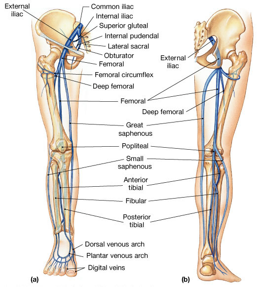
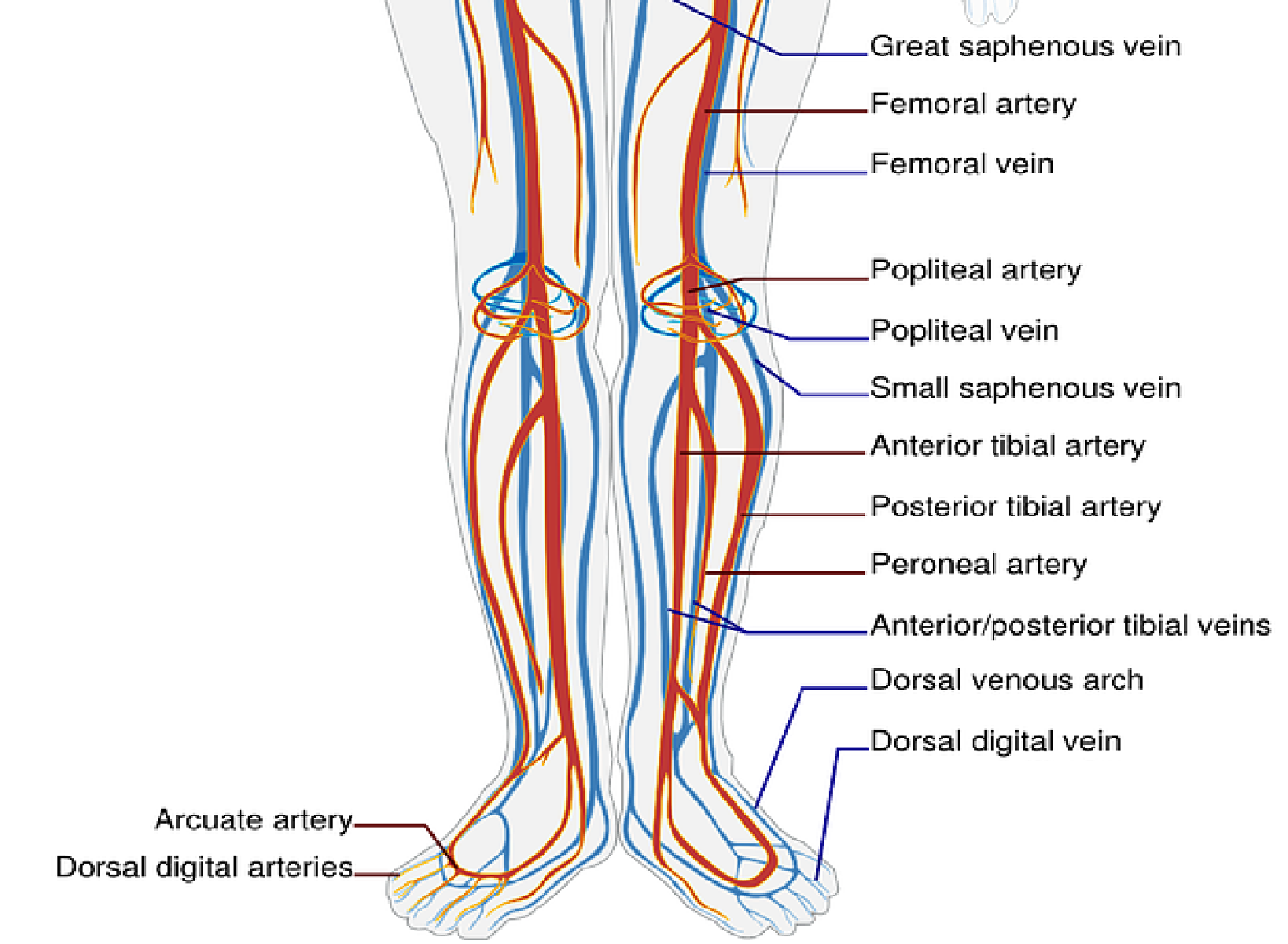
The popliteal vein enters the thigh through the adductor canal. The popliteal vein becomes the femoral vein when it passes through the adductor hiatushunters canal along with the artery and the saphenous nerve a branch of the femoral nerve. In the leg the anterior tibial posterior tibial peroneal and gastrocnemius veins join forming the popliteal vein at the popliteal fossa ascending into the thigh and becoming the femoral vein.
In one study Kubik 1985 in Appendix 2 a single popliteal trunk vein was found in the popliteal fossa in only 55 of legs. Once the popliteal vein has entered the thigh it is known as the femoral vein. The femoral vein becomes the 67.
The popliteal vein is one of the major blood vessels in the lower body. Interested in taking our award-winning Pocket Anatomy app for a test drive. In 40 there were two trunk veins which joined the femoral vein generally together to form a single femoral vein.
Rarely the popliteal vein runs more deeply to join. These arteries supply the myocardium. The deep vein of the thigh profunda.
The popliteal vein runs up through the popliteal fossa lying more posterior and usually medial to the artery. On the posterior aspect of the knee the anterior tibial posterior tibial and fibular veins join to form the popliteal vein. The calf veins join to form the popliteal vein or veins there may be two or sometimes three channels especially if there is a dual superficial femoral vein.
The arterial system has one of these the venous system has two Brachiocephalic These arteries supply the myocardium Coronary The more anterior artery pair serving the brain. Anterior and posterior tibial veins. After passing through the knee area the popliteal vein becorm 66.
It ascends through the popliteal fossa and becomes the femoral vein. Two veins that join forming the popliteal vein. Once the femoral vein passes the inguinal ligament it is renamed the external iliac vein.
Th e vein unites at the knee with the vein to form the popliteal vein. Major artery serving the skin and scalp of the head. At the opening in the adductor Magnus the vein continues as the femoral vein.
Two gastrocnemius veins pour into the same lateral vein of the popliteal vein medialis to. The cephalic vein and the axillary vein. Two veins that join forming the popliteal vein Anterior tibial and posterior tibial Artery generally used to take the pulse at the wrist Radial.
Arterial system has one of these the venous system has two. The short saphenous vein pierces the deep fascia of the leg and enters the popliteal fossa by passing through the two heads of. The popliteal vein starts at the lower border of the popliteus where veins accompanying the anterior and posterior tibial arteries join.
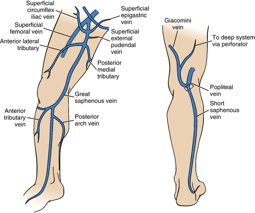
It is situated anteriorly accompanying the femoral artery. These two veins join with the subclavian vein to form the innominate vein. It is joined by the lesser saphenous vein from the superficial system ascends and forms the superficial femoral vein and ultimately the common femoral vein.
However in around 2 of cases duplicate trunk veins join duplicate femoral veins. The right common iliac vein and the left common iliac vein join to form the 19. The fibular vein drains the lateral portion of the leg and joins with the popliteal vein at the knee to form the.
Formed on the posterior aspect of the leg as the two tibial veins merge. Which two veins unite at the knee to form the popliteal vein. The popliteal vein runs up through the popliteal fossa lying more posterior and usually medial to the artery.
They join inferior to the knee to form the popliteal vein. The posterior tibial vein follows the posterior tibial artery penetrating the leg posteriorly to the medial malleolus of the ankle. The small saphenous sah-fe-nus vein drains the superficial posterior part of the leg and merges with the popliteal vein.
Accessory cephalic vein Anterior tibial vein. Artery generally used to take the pulse at the wrist. The popliteal vein enters the thigh via the adductor canal.
Two superficial veins in the arm. The cephalic vein is located laterally in the upper arm. The posterior tibial anterior tibial and common fibular veins.
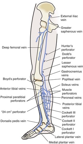
The connecting form of the three veins receiving blood flow from the sole of the foot peroneal vein posterior tibial vein and anterior tibial vein is gradual one vein joining at a time and thus progressively developing to the popliteal trunk. As well as the veins from the calf and calf muscles it is joined by the small saphenous vein at the saphenopopliteal junction. These veins join to form the posterior tibial and fibular veins.
It also receives venous blood from the superficial vein of the lateral leg ie. Anatomy and Physiology questions and answers. The popliteal vein becomes the femoral vein at the upper border of the popliteal fossa.
The longest vein in the body is the 21. Anterior tibial posterior tibial. In the groin the femoral vein is joined by the deep femoral vein which enters the abdomen underneath the inguinal ligament becoming the external iliac vein 1.
The anterior and posterior tibial veins drain the foot and deep regions of the leg. Give 3 examples of. 2 veins that join forming the popliteal vein.
It runs medial to the popliteal artery in the lower fossa posterior to it in the center and posterolateral to it in the upper fossa. The short saphenous vein. The popliteal vein is formed by the confluence of the deep veins of the leg ie.
On the posterior surface of the knee the anterior tibial posterior tibial and fibular veins unite to form the popliteal vein. The dural venous sinuses are drained by the 23.
![]()
Popliteal Vein Anatomy And Location Kenhub

Pin On Work
Femoral And Popliteal Vein Pocus A The Illustration Shows The Two Download Scientific Diagram

Anatomy Of Deep Venous System A And The Superficial Veins B Download Scientific Diagram
2
Venous Disease Thoracic Key

Arteries And Veins Of The Lower Limb Ppt Download
2

Pathophysiology Nursing School Survival Superficial Veins Medical Ultrasound

Arteries And Veins Of The Lower Limb Ppt Download
Anatomy Bony Pelvis And Lower Limb Foot Veins Article
/GettyImages-87313663-16bdfeaf37d048dbaef06b4f00b269b5.jpg)
Popliteal Vein Anatomy And Function
![]()
Veins Of The Lower Limb Anatomy Kenhub
Lower Extremity Veins Radiology Key
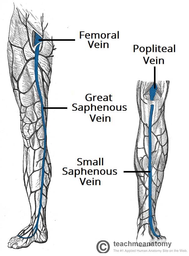
Venous Drainage Of The Lower Limb Teachmeanatomy

Ch 6 Peripheral Venous Evaluation Flashcards Quizlet
2

Pin En Fisiopatologia Insuficiencia Venosa
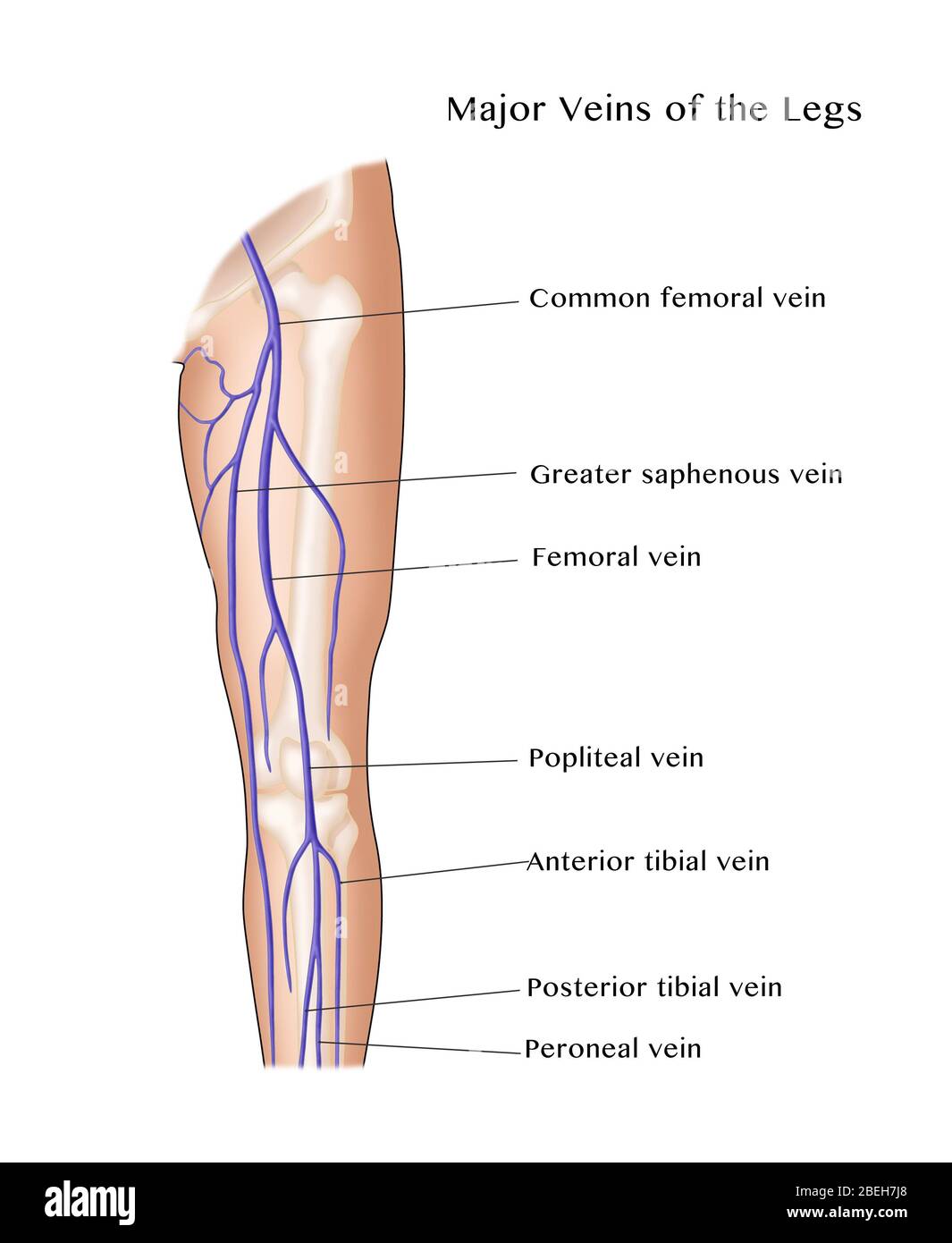
Major Veins High Resolution Stock Photography And Images Alamy


Comments
Post a Comment